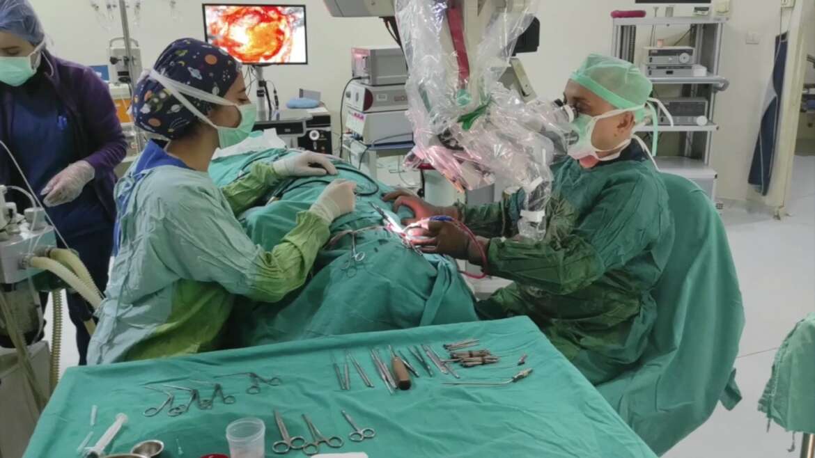
SURGERY FOR VERTIGO
The most common causes of vertigo are inner ear diseases. There are diseases related to the inner ear balance system that are not yet defined in classical books. We conduct research to find new treatments for our patients with our own knowledge, experience and experience. We can summarize the cases in which we apply surgical treatment.
- Eustachian Balloon Tuboplasty: Eustachian Tube is a canal that adjusts the pressure of the ear between the middle ear and the nasopharynx. Eustachian dysfunction is present in some inner ear balance system diseases. Especially in vertigo that does not go away with repetitive medical treatment and maneuvers, if there is eustachian canal dysfunction, it is a treatment that we apply and we get very effective results. At the same time, a long-lasting ventilation tube is attached to the ear. We get very satisfactory results in the prevention of attacks, which we also apply in Menire’s Disease, which is mentioned below.

- MENIERE’S DISEASE: It was first described by Prosper Ménierè in 1861. It is typically an inner ear disease with 4 main features; these are vertigo that occurs in attacks, sensorineural hearing loss (SNHL) with fluctuations and gradual progression, pressure sensation in the ear and tinnitus. Intratympanic treatments are applied to patients with recurrent severe attacks. Steroid injections are mostly administered intratympanically. However, if the attacks do not go away and the hearing is moderately severely impaired, intratympanic gentamicin treatment, which also destroys residual hearing, is started (Chemical labyrinthectomy). In this way, success is achieved in the treatment of vertigo, but the patient has permanent sensorineural hearing loss and tinnitus. In surgical treatments, endolymphatic sac decompression, endolymphatic sac tube application, vestibular neurectomy and ear ventilation tube application are procedures that preserve hearing. The success probability of invasive and non-invasive treatments is around 50-60%, and they are not superior to each other. Labrientectomy used to be for destruction, but nowadays the same results can be obtained with chemical labyrinthectomy.
- PERILYMPH FISTULA: Abnormal connection between the inner and middle ear and leakage of perilymph or endolymph fluid. Leakage may occur naturally through an oval or round window, or it may be caused by bone dehiscences. With sudden pressure changes, dizziness, hearing loss, tinnitus and nystagmus occur. fistula test; If the pressure in the ear canal is increased with a pneumatic otoscope, vertigo and nystagmus occur in the patient. Treatment; bed rest, elevation of the patient’s head, laxatives, anti-vertiginous drugs are prescribed. Unresponsive cases require surgical exploration. Often, leaks cannot be detected and possible locations are obliterated surgically

- TEMPORAL FRACTURES: Blunt traumas and transverse fractures of the temporal bone may occur if they pass through the vestibule or iatrogenically after mastoidectomy and stapes surgery.
- OTITIS MEDIA: Cholestetatomas in chronic otitis media can cause labyrinthitis. Labyrinthitis is of two classes, serous or suppurative. Serous labyrinthitis occurs when bacterial toxins affect the inner ear. It is manifested by sudden and high frequency hearing loss. Suppurative labyrinthitis occurs when bacteria directly affect the inner ear. Severe vestibular and cochlear symptoms occur and permanent sequelae occur. In chronic otitis media with cholesteatoma, vertigo may occur if bone erosion occurs on the semicircular canal or vestibule. Mastoidectomy and obliteration of pathology are performed.kemik erozyonu oluşursa, vertigo oluşabilir. Mastoidektomi ve patolojinin obliterasyonu yapılır.

- TUMORS: Tumors seen in the pontocerebellar corner affect the vestibular nerve and cause vertigo. Acoustic Neuromas are seen last. The most common symptom is unilateral hearing loss. Tinnitus and dizziness may accompany symptoms. Hearing loss usually occurs at high frequencies (4-8 kHz). Even small tumors can be visualized by MRI with gadolinium. It most often develops from the superior branch of the vestibular nerve. Since it grows slowly (2.5-4 mm/year), its symptoms are not obvious. When the tumor reaches a certain size (>2.5 cm), the facial and trigeminal nerves are involved. Tumor growth is stopped with cyberkinfe, gama knife, or steortactic radiosurgery. If progressive and compression symptoms occur, surgical treatment is required..


